Rubie Rounkles
  • Blog
  • About
  • Contact

Leg Length Discrepancy Weight Lifting

7/2/2017

 
Overview

Limb deformity or leg length problems can be treated by applying an external frame to the leg. The frame consists of metal rings which go round the limb. The rings are held onto the body by wires and metal pins which pass through the skin and are anchored into the bone. During this operation, the bone is divided. Gradual adjustment of the frame results in creation of a new bone allowing a limb to be lengthened. The procedure involves the child having an anaesthetic. The child is normally in hospital for one week. The child and family are encouraged to clean pin sites around the limb. The adjustments of the frame (distractions) are performed by the child and/or family. The child is normally encouraged to walk on the operated limb and to actively exercise the joints above and below the frame. The child is normally reviewed on a weekly basis in clinic to monitor the correction of the deformity. The frame normally remains in place for 3 months up to one year depending on the condition which is being treated. The frame is normally removed under a general anaesthetic at the end of treatment.Leg Length Discrepancy

Causes

Sometimes the cause of LLD is unknown, yet the pattern or combination of conditions is consistent with a certain abnormality. Examples include underdevelopment of the inner or outer side of the leg (hemimelias) or (partial) inhibition of growth of one side of the body of unknown cause (hemihypertrophy). These conditions are present at birth, but the limb length difference may be too small to be detected. As the child grows, the LLD increases and becomes more noticeable. In hemimelia, one of the two bones between the knee and the ankle (tibia or fibula) is abnormally short. There also may be associated foot or knee abnormalities. Hemihypertrophy or hemiatrophy are rare conditions in which there is a difference in length of both the arm and leg on only one side of the body. There may also be a difference between the two sides of the face. Sometimes no cause can be found. This type of limb length is called idiopathic. While there is a cause, it cannot be determined using currect diagnostic methods.

Symptoms

Back pain along with pain in the foot, knee, leg and hip on one side of the body are the main complaints. There may also be limping or head bop down on the short side or uneven arm swinging. The knee bend, hip or shoulder may be down on one side, and there may be uneven wear to the soles of shoes (usually more on the longer side).

Diagnosis

There are several orthopedic tests that are used, but they are rudimentary and have some degree of error. Even using a tape measure with specific anatomic landmarks has its errors. Most leg length differences can be seen with a well trained eye, but I always recommend what is called a scanagram, or a x-ray bone length study (see picture above). This test will give a precise measurement in millimeters of the length difference.

Non Surgical Treatment

In some circumstances, the physician will recommend a non-surgical form of treatment. Non-surgical treatments include orthotics and prosthetics. Orthotics are a special type of lift placed in or on a shoe that can be used in the treatment of leg length discrepancies between two and six centimeters. In pediatric patients who have large discrepancies and are not good candidates for other treatment forms, prosthetics can be helpful.

LLL Shoe Insoles

how to increase height in 1 week

Surgical Treatment

Surgery is another option. In some cases the longer extremity can be shortened, but a major shortening may weaken the muscles of the extremity. In growing children, lower extremities can also be equalized by a surgical procedure that stops the growth at one or two sites of the longer extremity, while leaving the remaining growth undisturbed. Your physician can tell you how much equalization can be attained by surgically halting one or more growth centers. The procedure is performed under X-ray control through very small incisions in the knee area. This procedure will not cause an immediate correction in length. Instead, the LLD will gradually decrease as the opposite extremity continues to grow and "catch up." Timing of the procedure is critical; the goal is to attain equal length of the extremities at skeletal maturity, usually in the mid- to late teens. Disadvantages of this option include the possibility of slight over-correction or under-correction of the LLD and the patient?s adult height will be less than if the shorter extremity had been lengthened. Correction of significant LLDs by this method may make a patient?s body look slightly disproportionate because of the shorter legs.
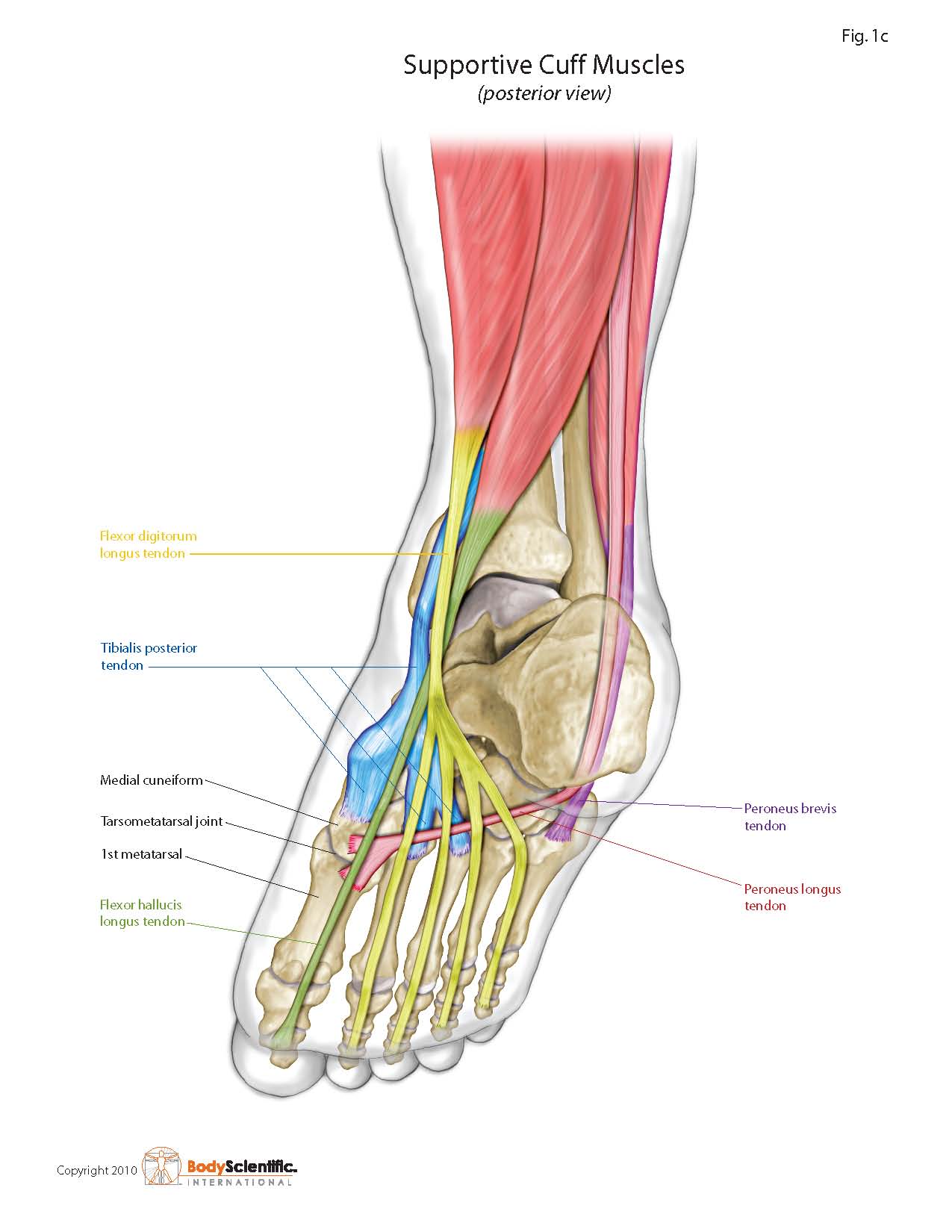
Ways To Diagnose Fallen Arches

7/2/2017

 
Overview

Acquired Flat Feet

Over-pronation is very common and affects millions of Australians. To better understand this condition, we'll take a closer look at the 3 most common foot types. An estimated 70% of the population has fallen arches (or a low arch). Only 20% has a normal arch. And 10% have abnormal feet, in other words they either have flat feet or the opposite - a high arched foot.

Causes

As discussed above, many health conditions can create a painful flatfoot. Damage to the posterior tibial tendon is the most common cause of AAFD. The posterior tibial tendon is one of the most important tendons of the leg. It starts at a muscle in the calf, travels down the inside of the lower leg and attaches to the bones on the inside of the foot. The main function of this tendon is to hold up the arch and support your foot when you walk. If the tendon becomes inflamed or torn, the arch will slowly collapse. Women and people over 40 are more likely to develop problems with the posterior tibial tendon. Other risk factors include obesity, diabetes, and hypertension. Having flat feet since childhood increases the risk of developing a tear in the posterior tibial tendon. In addition, people who are involved in high impact sports, such as basketball, tennis, or soccer, may have tears of the tendon from repetitive use.

Symptoms

Most people do not exhibit any symptoms of flat feet, but if the condition is due to an underlying problem, symptoms can include foot pain, mainly in the arch or heel areas, difficulty standing on tiptoes, swelling that occurs on the inside of the ankle, pain in the calf, knee, hip, or lower leg area, both feet lie flat on the ground with no gap, Shoes may not fit properly, heel may tilt away from the midline of the body more than usual, absence of foot arch when standing. If you are experiencing these symptoms and have flat feet, you should consider seeing your doctor or a podiatrist immediately for an examination.

Diagnosis

People who have flat feet without signs or symptoms that bother them do not generally have to see a doctor or podiatrist about them. However, if any of the following occur, you should see your GP or a podiatrist. The fallen arches (flat feet) have developed recently. You experience pain in your feet, ankles or lower limbs. Your unpleasant symptoms do not improve with supportive, well-fitted shoes. Either or both feet are becoming flatter. Your feet feel rigid (stiff). Your feet feel heavy and unwieldy. Most qualified health care professionals can diagnose flat feet just by watching the patient stand, walk and examining his/her feet. A doctor will also look at the patient's medical history. The feet will be observed from the front and back. The patient may be asked to stand on tip-toe while the doctor examines the shape and functioning of each foot. In some cases the physician may order an X-ray, CT (computed tomography) scan, or MRI (magnetic resonance imaging) scan.

How do you get an arch in your foot?

Non Surgical Treatment

Treatment often consists of giving the affected foot support from underneath so that the strain is taken off it (by the use of insoles or support pads) and so symptoms are relieved. A specialist in this problem is known as a podiatrist and these do work in the NHS - ask you father's doctor whether his condition is bad enough to warrant a referral to such a specialist.

Surgical Treatment

Flat Feet

Fallen arches may occur with deformities of the foot bones. Tarsal coalition is a congenital condition in which the bones of the foot do not separate from one another during development in the womb. A child with tarsal coalition exhibits a rigid flat foot, which can be painful, notes the patient information website eOrthopod. Surgery may prove necessary to separate the bones. Other foot and ankle conditions that cause fallen arches may also require surgery if noninvasive treatments fail to alleviate pain and restore normal function.

Prevention

Donning a first-rate pair of arch supports, therapeutic socks and proper footwear before heading out to enjoy hours of holiday fun is one option to consider. Your podiatrist can help you find just the right ones. Once you have them on, they?ll help ease the amount of pressure being put on your body and keep the blood flowing in the right direction. While you?re standing in line, consider doing a bit of exercise as well. We?re not talking about channeling your inner Jack LaLanne here. Otherwise, you might attract the attention of the mall security guards. Simple ankle rotations and walking in place may help to reduce edema and give your flat feet a bit of a break. If you happen to be in a shopping mall or center where foot massages are available, take advantage of them periodically. They are likely to make you feel better and it?s a great excuse to carve out a few quiet moments for yourself. If you can?t visit a professional, tuck a personal foot massager into your purse. That way, you can lightly massage your own feet during the car ride home. Lastly, there are certain foods and nutritional supplements available that may reduce edema caused by standing on flat feet for hours at a time. The list includes potassium rich foods like raisins, bananas, baby carrots, nuts and yogurt. So, you may want to pack a snack for those trips to the mall or hit the food court before you hit the stores.

After Care

Time off work depends on the type of work as well as the surgical procedures performed. . A patient will be required to be non-weight bearing in a cast or splint and use crutches for four to twelve weeks. Usually a patient can return to work in one to two weeks if they are able to work while seated. If a person's job requires standing and walking, return to work may take several weeks. Complete recovery may take six months to a full year. Complications can occur as with all surgeries, but are minimized by strictly following your surgeon's post-operative instructions. The main complications include infection, bone that is slow to heal or does not heal, progression or reoccurrence of deformity, a stiff foot, and the need for further surgery. Many of the above complications can be avoided by only putting weight on the operative foot when allowed by your surgeon.

All You Ought To Understand About

6/28/2017

 
Overview

Heel Pain

Pain arises in the centre of the heel and sometimes can spread to the arch region of the foot. Some patients complain of intense pain when walking the first few steps in the morning and after periods of rest. The most common cause of heel pain is plantar fascitis. The plantar fascia is a broad band of tissue that runs along the bottom of the foot. When this band of tissue is damaged due to biomechanical reasons or inappropriate running shoes then the tissue can become inflamed or even tear at the heel bone. This can be very painful.

Causes

Plantar Fasciitis is the most common form of heel pain. The tears and inflammation that develop along the plantar fascia ligament result in dull aching pain or a burning sensation along the bottom of the foot. Pain becomes particularly noticeable after periods of rest, such as during the first few steps after getting out of bed in the morning, or after getting up after a prolonged period of sitting. Another common form of heel pain is the development of a heel spur. A heel spur, as mentioned above, is the formation of a bony hook extending from the heel. Typically, these growths develop near the area where the plantar fascia connects to the heel bone. The repetitive pressure on the plantar fascia that results from stretching excessively away from the heel bone causes a response from our body that delivers calcium to the area. The heel pain that ensues develops from the nerves and sensitive tissue that become irritated when the bone fragment digs into the bottom of the heel. Pain may decrease after walking as the tissue in the heel gets used to the fragment and adjusts around it. However, pain will be particularly problematic following periods of rest. Strained muscle tissue may cause heel pain in several areas. A tight plantar fascia causes additional tension, particularly while exercising, placing runners and other athletes at risk if the ligament is not properly warmed up prior to exercise. Additionally, a tight Achilles tendon along the back of the foot can also add tension along the plantar fascia, resulting in possible damage, not to mention the damage and pain that can occur along the Achilles tendon itself (Achilles tendonitis). It is recommended that athletes properly stretch the foot as well as the calf in order to reduce tension on muscle and other tissue in the foot.

Symptoms

Initially, this pain may only be present when first standing up after sleeping or sitting. As you walk around, the muscle and tendon loosen and the pain goes away. As this problem progresses, the pain can be present with all standing and walking. You may notice a knot or bump on the back of the heel. Swelling may develop. In some cases, pressure from the back of the shoe causes pain.

Diagnosis

To arrive at a diagnosis, the foot and ankle surgeon will obtain your medical history and examine your foot. Throughout this process the surgeon rules out all the possible causes for your heel pain other than plantar fasciitis. In addition, diagnostic imaging studies such as x-rays or other imaging modalities may be used to distinguish the different types of heel pain. Sometimes heel spurs are found in patients with plantar fasciitis, but these are rarely a source of pain. When they are present, the condition may be diagnosed as plantar fasciitis/heel spur syndrome.

Non Surgical Treatment

Treatment for plantar fasciitis - the vast majority of patients recover with conservative treatments (designed to avoid radical medical therapeutic measures or operative procedures) within months. Heel with ice-pack. Home care such as rest, ice-pack use, proper-fitting footwear and foot supports are often enough to ease heel pain. Non-steroidal anti-inflammatory drugs (NSAIDs) - medications with analgesic (pain reducing), antipyretic (fever reducing) effects. In higher doses they also have anti-inflammatory effects, they reduce inflammation (swelling). Non-steroidal distinguishes NSAIDs from other drugs which contain steroids, which are also anti-inflammatory. NSAIDs are non-narcotic (they do not induce stupor). For patients with plantar fasciitis they may help with pain and inflammation. Corticosteroids, a corticosteroid solution is applied over the affected area on the skin; an electric current is used to help absorption. Alternatively, the doctor may decide to inject the medication. However, multiple injections may result in a weakened plantar fascia, significantly increasing the risk of rupture and shrinkage of the fat pad covering the heel bone. Some doctors may use ultrasound to help them make sure they have injected in the right place Corticosteroids are usually recommended when NSAIDs have not helped. Physical therapy, a qualified/specialized physical therapist (UK: physiotherapist) can teach the patient exercises which stretch the plantar fascia and Achilles tendon, as well as strengthening the lower leg muscles, resulting in better stabilization of the ankle and heel. The patient may also be taught how to apply athletic taping, which gives the bottom of the foot better support. Night splints, the splint is fitted to the calf and foot; the patient keeps it on during sleep. Overnight the plantar fascia and Achilles tendon are held in a lengthened position; this stretches them. Orthotics, insoles and orthotics (assistive devices) can be useful to correct foot faults, as well as cushioning and cradling the arch during the healing process. Extracorporeal shock wave therapy, sound waves are aimed at the affected area to encourage and stimulate healing. This type of therapy is only recommended for chronic (long-term) cases, which have not responded to conservative therapy.

Surgical Treatment

If treatment hasn't worked and you still have painful symptoms after a year, your GP may refer you to either an orthopaedic surgeon, a surgeon who specialises in surgery that involves bones, muscles and joints or a podiatric surgeon, a podiatrist who specialises in foot surgery. Surgery is sometimes recommended for professional athletes and other sportspeople whose heel pain is adversely affecting their career. Plantar release surgery is the most widely used type of surgery for heel pain. The surgeon will cut the fascia to release it from your heel bone and reduce the tension in your plantar fascia. This should reduce any inflammation and relieve your painful symptoms. Surgery can be performed either as open surgery, where the section of the plantar fascia is released by making a cut into your heel or endoscopic or minimal incision surgery - where a smaller incision is made and special instruments are inserted through the incision to gain access to the plantar fascia. Endoscopic or minimal incision surgery has a quicker recovery time, so you will be able to walk normally much sooner (almost immediately), compared with two to three weeks for open surgery. A disadvantage of endoscopic surgery is that it requires both a specially trained surgical team and specialised equipment, so you may have to wait longer for treatment than if you were to choose open surgery. Endoscopic surgery also carries a higher risk of damaging nearby nerves, which could result in symptoms such as numbness, tingling or some loss of movement in your foot. As with all surgery, plantar release carries the risk of causing complications such as infection, nerve damage and a worsening of your symptoms after surgery (although this is rare). You should discuss the advantages and disadvantages of both techniques with your surgical team.

heel bursitis

Prevention

Feet Pain

You can reduce the risk of heel pain in many ways, including. Wear shoes that fit you properly with a firm fastening, such as laces. Choose shoes with shock-absorbent soles and supportive heels. Repair or throw out any shoes that have worn heels. Always warm up and cool down when exercising or playing sport, include plenty of slow, sustained stretches. If necessary, your podiatrist will show you how to tape or strap your feet to help support the muscles and ligaments. Shoe inserts (orthoses) professionally fitted by your podiatrist can help support your feet in the long term.

What Is Mortons Neuroma

5/30/2017

 
Overview

interdigital neuromaMorton's neuroma is named after Dr Morton who first described this condition in 1876. It is sometimes called Morton's metatarsalgia or interdigital neuroma. It is a condition that affects one of the common plantar digital nerves that run between the long bones (metatarsals) in the foot. It most commonly affects the nerve between the third and fourth metatarsal bones, causing pain and numbness in the third and fourth toes. It can also affect the nerve between the second and third metatarsal bones, causing symptoms in the second and third toes. Morton's neuroma rarely affects the nerve between the first and second, or between the fourth and fifth, metatarsal bones. It tends to affect only one foot. It is rare to get two neuromas at the same time in the same foot.

Causes

The source of this pain is an enlargment of the sheath of an intermetatarsal nerve in the foot. This usually occurs in the third intermetatarsal space, the space between the third and fourth toes and metatarsals. It occurs here, at the site third intermetatarsal nerve, since this intermetatarsal nerve is the thickest being comprised of the joining of two different nerves. It also may occur in the other intermetatarsal areas, with the second interspace being the next most common location.

Symptoms

Feelings of numbness, tingling or tenderness in the ball of the foot (the area just behind the base of the toes) are some of the first signs of a condition known as Morton?s Neuroma. However, the condition is somewhat unpredictable, and symptoms may vary from patient to patient. Generally, however, the discomfort gets worse rather than better, and the patient may feel pain or a burning sensation that radiates out to the toes. Eventually, wearing shoes becomes uncomfortable (or even unbearable), and the patient may complain that the feeling is similar to that of having a stone bruise, or walking on a marble or pebble constantly, even though no there is no trauma to the skin, and no visible bump or lump on the sole of the foot.

Diagnosis

The clinical symptoms should quickly lead your doctor to suspect a neuroma. When examined, the doctor may feel a "click" which is known as Mulder's sign. There may be tenderness in the interspace. The metatarsal bones will also be examined both clinically (and often with an xray). Tenderness at one of the metatarsal bones can suggest an overstress reaction (pre-stress fracture or stress fracture) in the bone. An ultrasound scan can confirm the diagnosis and is a less expensive and at this time, at least as sensitive a test as an MRI. An x-ray does not show neuromas, but can be useful to "rule out" other causes of the pain.

Non Surgical Treatment

You may be able to treat this problem at home. Avoid wearing tight, pointy, or high-heeled shoes. Choose well-fitted shoes with plenty of room for your toes. Put ice or a cold pack on the area for 10 to 15 minutes at a time. Put a thin cloth between the ice and your skin. Take anti-inflammatory medicines to reduce pain and swelling. These include ibuprofen (such as Advil or Motrin) and naproxen (such as Aleve). Rest your feet when you can. Reduce activities that put pressure on the toes, such as racquet sports or running. Try massaging your foot to relax the muscles around the nerve. If these steps do not relieve your symptoms, your doctor may have you use special pads or devices that spread the toes to keep them from squeezing the nerve. In some cases, a doctor may give a steroid shot to reduce swelling and pain. If these treatments do not help, your doctor may suggest surgery.plantar neuroma

Surgical Treatment

Surgery for neuroma most often involves removing affected nerve in the ball of the foot. An incision is made on the top of the foot and the nerve is carefully removed. Surgeon must remove the nerve far enough back so that the nerve doesn?t continue to become impinged at the ball of the foot. Alternatitvely, another type of surgery involves releasing a tight ligament that encases the nerve. Recovery after Morton?s neuroma (neurectomy) surgery is generally quick. Typically patients are walking on the operated foot in a post-surgical shoe for 2 - 4 weeks, depending on healing. Return to shoes is 2-6 weeks after the surgery. Factors that may prolong healing are age, smoking, poor nutritional status, and some medical problems.

The Ideal Solution To Leg Length Discrepancy Are Shoe Lifts

2/23/2016

 
There are actually not one but two different types of leg length discrepancies, congenital and acquired. Congenital means you are born with it. One leg is structurally shorter compared to the other. As a result of developmental stages of aging, the human brain picks up on the gait pattern and recognizes some difference. The entire body typically adapts by tilting one shoulder to the "short" side. A difference of less than a quarter inch is not grossly excessive, demand Shoe Lifts to compensate and ordinarily won't have a profound effect over a lifetime.

Leg Length Discrepancy Shoe Lift

Leg length inequality goes largely undiscovered on a daily basis, however this issue is very easily solved, and can eliminate a number of cases of upper back pain.

Therapy for leg length inequality commonly consists of Shoe Lifts . These are economical, regularly being under twenty dollars, in comparison to a custom orthotic of $200 or maybe more. When the amount of leg length inequality begins to exceed half an inch, a whole sole lift is generally the better choice than a heel lift. This prevents the foot from being unnecessarily stressed in an abnormal position.

Upper back pain is easily the most common ailment impacting men and women today. Over 80 million men and women experience back pain at some point in their life. It is a problem which costs companies vast amounts of money each year as a result of time lost and output. Innovative and superior treatment solutions are continually sought after in the hope of decreasing the economical impact this issue causes.

Leg Length Discrepancy Shoe Lifts

People from all corners of the world suffer the pain of foot ache due to leg length discrepancy. In a lot of these cases Shoe Lifts might be of very useful. The lifts are capable of reducing any discomfort and pain in the feet. Shoe Lifts are recommended by many expert orthopaedic practitioners".

In order to support the human body in a well balanced fashion, the feet have got a critical function to play. Irrespective of that, it is often the most neglected zone in the body. Some people have flat-feet meaning there may be unequal force placed on the feet. This causes other parts of the body including knees, ankles and backs to be affected too. Shoe Lifts ensure that correct posture and balance are restored.

What Is A Posterior Calcaneal Spur

9/28/2015

 
Inferior Calcaneal Spur

Overview

If you suffer from heel pain, you know that it affects every step you take. And by the time you take off your shoes in the evening, your feet are really suffering. The pain you?re feeling could be caused by heel bone spurs. Heel bone spurs are common in people who walk, stand or run on hard surfaces such as concrete or tile floors-and that?s most of us! This kind of frequent, intense impact on hard surfaces overstretches and can even tear the ligaments on the bottom of the foot, a condition known as plantar fasciitis. In extreme cases, these ligaments begin to pull away from the bone. Heel bone spurs are created because of this injury to the foot. They are not painful by themselves, but they do irritate surrounding tissues, which causes heel pain.

Causes

The cause of heel spurs is excessive strain placed on the plantar fascia over a long period of time, as a result of different factors. These factors include incorrect gait, being overweight, ageing or being in a job that requires a lot of standing on hard floors. It is usually a combination of any of these factors that will bring on the development of heel spurs.

Calcaneal Spur

Symptoms

More often than not, heel spurs have no signs or symptoms, and you don?t feel any pain. This is because heel spurs aren?t pointy or sharp pieces of bone, contrary to common belief. Heel spurs don?t cut tissue every time movement occurs; they?re actually deposits of calcium on bone set in place by the body?s normal bone-forming mechanisms. This means they?re smooth and flat, just like all other bones. Because there?s already tissue present at the site of a heel spur, sometimes that area and the surrounding tissue get inflamed, leading to a number of symptoms, such as chronic heel pain that occurs when jogging or walking.

Diagnosis

The proper diagnosis of a heel spur often requires an X-ray. To make this process as convenient for his patients as possible, most clinics have an on-site digital X-ray and diagnostic ultrasound machines. This can make it unnecessary for patients to visit diagnostic imaging centers, allowing patients to receive more expedient treatment.

Non Surgical Treatment

There are many temporary solutions to resolve the pain associated with irritation to the plantar ligaments. Common recommendations are ice and anti-inflammatory medications or even cortisone injections, however none of these solve the fundamental problem. To permanently resolve heel spurs you need to support and restrict the movement of the plantar ligaments. Flexible shoes will aggravate and often contribute to heel spurs. We recommend a RIGID orthotic that extends from the metatarsal heads to the heel to resolve heel spurs.

Surgical Treatment

Almost 90% of the people suffering from heel spur get better with nonsurgical treatments. However, if the conservative treatments do not help you and you still have pain even after 9 to 12 months, your doctor may advise surgery for treating heel spur. The surgery helps in reducing the pain and improving your mobility. Some of the surgical techniques used by doctors are release of the plantar fascia. Removal of a spur. Before the surgery, the doctor will go for some pre-surgical tests and exams. After the operation, you will need to follow some specific recommendations which may include elevation of the foot, waiting time only after which you can put weight on the foot etc.

Prevention

o help prevent heel and bone spurs, wear properly designed and fitted shoes or boots that provide sufficient room in the toe box so as not to compress the toes. They should also provide cushioning in appropriate areas to minimize the possibility of the irritation and inflammation that can lead to bone spurs in the feet. If needed, use inserts that provide arch support and a slight heel lift to help ensure that not too much stress is placed on the plantar fascia. This helps to reduce the possibility of inflammation and overstress. Wearing padded socks can also help by reducing trauma. Peer-reviewed, published studies have shown that wearing clinically-tested padded socks can help protect against injuries to the skin/soft tissue of the foot due to the effects of impact, pressure and shear forces. Also consider getting your gait analyzed by a foot health professional for appropriate orthotics. If you have heel pain, toe pain or top-of-the-foot pain, see your doctor or foot specialist to ensure that a spur has not developed.

Inferior Calcaneal Spur Symtoms

9/27/2015

 
Posterior Calcaneal Spur

Overview

Heel spurs are a common foot problem resulting from excess bone growth on the heel bone. The bone growth is usually located on the underside of the heel bone, extending forward to the toes. One explanation for this excess production of bone is a painful tearing of the plantar fascia connected between the toes and heel. This can result in either a heel spur or an inflammation of the plantar fascia, medically termed plantar fascitis. Because this condition is often correlated to a decrease in the arch of the foot, it is more prevalent after the age of six to eight years, when the arch is fully developed.

Causes

A strong band of sinew (plantar fascia) stretches across the sole of the foot below the surface of the skin and is attached to a point in the middle of the under surface of the heel bone. With repeated activity on our feet, the plantar fascia can become tight and cause persistent traction (tugging) on its attachment point into the heel bone, and inflammation and pain may develop at this site. This painful condition is known as plantar fasciitis. Sometimes a ?spur? develops at the site of this traction on the bone and protrudes into the surrounding tissue. This is a heel spur.

Calcaneal Spur

Symptoms

More often than not, heel spurs have no signs or symptoms, and you don?t feel any pain. This is because heel spurs aren?t pointy or sharp pieces of bone, contrary to common belief. Heel spurs don?t cut tissue every time movement occurs; they?re actually deposits of calcium on bone set in place by the body?s normal bone-forming mechanisms. This means they?re smooth and flat, just like all other bones. Because there?s already tissue present at the site of a heel spur, sometimes that area and the surrounding tissue get inflamed, leading to a number of symptoms, such as chronic heel pain that occurs when jogging or walking.

Diagnosis

A thorough medical history and physical exam by a physician is always necessary for the proper diagnosis of heel spurs and other foot conditions. X rays of the heel area are helpful, as excess bone production will be visible.

Non Surgical Treatment

Only in rare cases do the symptoms of heel spurs fail to be resolved through conservative treatment. Conservative treatment, although not 100% effective, is successful in most cases and should be given ample time to work. In many cases, conservative methods should be utilized as long as a year depending on the rate at which your body responds to the treatment. When treatment is unsuccessful, surgery may be considered. A common surgical procedure for this condition is plantar fascia release surgery. In this procedure, the tension of the plantar fascia ligament is released, lessening tension in the heel and helping to prevent damage.

Surgical Treatment

Most studies indicate that 95% of those afflicted with heel spurs are able to relieve their heel pain with nonsurgical treatments. If you are one of the few people whose symptoms don?t improve with other treatments, your doctor may recommend plantar fascia release surgery. Plantar fascia release involves cutting part of the plantar fascia ligament in order to release the tension and relieve the inflammation of the ligament. Sometimes the bone spur is also removed, if there is a large spur (remember that the bone spur is rarely a cause of pain. Overall, the success rate of surgical release is 70 to 90 percent in patients with heel spurs. One should always be sure to understand all the risks associated with any surgery they are considering.

Bursitis Of The Foot Therapy

8/23/2015

 
Overview

Bursitis occurs when a bursa is irritated from frequent pressure and it becomes inflamed. Due to the location close the Achilles tendon, Achilles bursitis is often mistaken for tendinitis. Achilles bursitis is a common overuse injury in runners, ice skaters and other athletes.

Causes

Retrocalcaneal bursitis can be caused through injury or infection or be can be triggered by certain health conditions. If bursitis develops as a result of injury then it will normally be due to a repetitive strenuous activity that encourages the calf muscles (the gastrocnemius and soleus muscle), which attach to the heel bone via the Achilles tendon, to tighten and shorten from overuse, for example repetitively wearing high heels, running and even wearing tight shoes that pinch at the back of the heel. This puts more pressure over the bursa as the tendon rubs more tightly over it, irritating it and triggering a painful inflammatory reaction (swelling). This risk of developing bursitis in this way is greater for those whose jobs or hobbies involve a lot of repetitive movements, for example carpet fitters and gardeners who spend a lot of time kneeling and so are more at risk of bursitis in the knee. Runners have a greater likelihood of developing bursitis in the hip. Bursitis can also be brought on by excessive pressure or direct impact trauma, such as banging your elbow or dropping on to your knees. Infection is a less common cause of bursitis and normally only occurs in people who have a weakened immune system from other health issues. The infection can work its way to the bursa from a cut close to the bursa that has become infected, in these cases the bursitis is termed as septic bursitis. Certain health conditions can also trigger the development of bursitis, such as rheumatoid arthritis and gout, amongst others.

Symptoms

Some of the symptoms of bursitis in the heel, or retrocalcaneal bursitis, are as described below. Severe pain in the heel area of the foot, sometimes radiating to the ankle, associated with physical activities like walking, jogging and even on physical contact to the area. The physical signs of heel bursitis, which are noticeable in the heel area, are reddish discoloration of the skin that is warm to touch.

Diagnosis

The diagnosis is based on the symptoms and an examination. For anterior Achilles tendon bursitis, doctors use x-rays to rule out a fracture of the heel bone or damage to the heel bone caused by rheumatoid arthritis or other inflammatory arthritis.

Non Surgical Treatment

Gradually progressive stretching of the Achilles tendon may help to relieve impingement on the subtendinous calcaneal bursa. Stretching of the Achilles tendon can be performed by having the patient place the affected foot flat on the floor and lean forward toward the wall until a gentle stretch is felt in the ipsilateral Achilles tendon. The stretch is maintained for 20-60 seconds and then is relaxed. Achilles stretch 1. The patient stands with the affected foot flat on the floor and leans forward toward the wall until a gentle stretch is felt in the ipsilateral Achilles tendon. The stretch is maintained for 20-60 seconds and then is relaxed. Achilles stretch 2. This stretch, which is somewhat more advanced than that shown in Images 1-2, isolates the Achilles tendon. It is held for at least 20-30 seconds and then is relaxed. To maximize the benefit of the stretching program, the patient should repeat the exercise for multiple stretches per set, multiple times per day. Ballistic (ie, abrupt, jerking) stretches should be avoided in order to prevent clinical exacerbation. The patient should be instructed to ice the posterior heel and ankle in order to reduce inflammation and pain. Icing can be performed for 15-20 minutes at a time, several times a day, during the acute period, which may last for several days. Some clinicians also advocate the use of contrast baths, ultrasound or phonophoresis, iontophoresis, or electrical stimulation for treatment of calcaneal bursitis. If the patient's activity level needs to be decreased as a result of this condition, alternative means of maintaining strength and cardiovascular fitness (eg, swimming, water aerobics) should be suggested.

Surgical Treatment

Surgery is rarely need to treat most of these conditions. A patient with a soft tissue rheumatic syndrome may need surgery, however, if problems persist and other treatment methods do not help symptoms.

Contracted Toe Causes

6/24/2015

 
Hammer ToeOverview

If the joint on one of your toes, usually the toe next to the big toe or the smallest toe, points upward rather than lying flat, you might have a hammertoe. The condition is actually a deformity that happens when one of the toe muscles becomes weak and puts pressure on the toe?s tendons and joints. This pressure forces the toe to become misshapen and stick up at the joint. Also, there?s frequently a corn or callus on top of the deformed toe. This outgrowth can cause pain when it rubs against the shoe.

Causes

Factors that may increase you risk of hammertoe and mallet toe include age. The risk of hammertoe and mallet toe increases with age. Your sex. Women are much more likely to develop hammertoe or mallet toe than are men. Toe length. If your second toe is longer than your big toe, it's at higher risk of hammertoe or mallet toe.

Hammer ToeSymptoms

A soft corn, or heloma molle, may exist in the web space between toes. This is more commonly caused by an exostosis, which is basically an extra growth of bone possibly due to your foot structure. As this outgrowth of excessive bone rubs against other toes, there is friction between the toes and a corn forms for your protection.

Diagnosis

The treatment options vary with the type and severity of each hammer toe, although identifying the deformity early in its development is important to avoid surgery. Your podiatric physician will examine and X-ray the affected area and recommend a treatment plan specific to your condition.

Non Surgical Treatment

Hammer toes may be effectively corrected in different ways. Treatments can be non-invasive and involve physical therapy along with the advice that the person not wear any more shoes that restrict appropriate space for their toes. Appropriate shoes for people who want to avoid hammer toes, or for people who already Hammer toe have them, should be at least half an inch longer than the person's longest toe. High-heeled shoes are something to definitely avoid.

Surgical Treatment

Surgery may be the treatment of choice if conservative approaches prove unsuccessful. Usually performed as an outpatient procedure, the specific surgery will depend on the type and extent of injury to the toe. Recovery my take several days or weeks and you may experience some redness, stiffness and swelling of the affected toe. Your physician will recommend taking it easy and to keep your foot elevated while you recover.

Hammer ToePrevention

elect and wear the right shoe for specific activities (such as running shoes for running). Alternate shoes. Don't wear the same pair of shoes every day. Avoid walking barefoot, which increases the risk for injury and infection. At the beach or when wearing sandals, always use sunblock on your feet, as you would on the rest of your body. Be cautious when using home remedies for foot ailments. Self-treatment can often turn a minor problem into a major one. It is critical that people with diabetes see a podiatric physician at least once a year for a checkup. People with diabetes, poor circulation, or heart problems should not treat their own feet, including toenails, because they are more prone to infection.

Over-Pronation

6/3/2015

 
Overview

When sitting, an over-pronating foot appears quite normal, i.e. showing a normal arch with room under the underside of the foot. The moment you get up and put weight on your feet the situation changes: the arches lower and the ankle slightly turns inwards. When you walk or run more weight is placed on the feet compared to standing and over-pronation will become more evident. When walking barefoot on tiles or timber floors over-pronation is more visible, compared to walking on carpet or grass.Pronation

Causes

Generally fallen arches are a condition inherited from one or both parents. In addition, age, obesity, and pregnancy cause our arches to collapse. Being in a job that requires long hours of standing and/or walking (e.g. teaching, retail, hospitality, building etc) contributes to this condition, especially when standing on hard surfaces like concrete floors. Last, but not least unsupportive footwear makes our feet roll in more than they should.

Symptoms

Common conditions seen with overpronation include heel pain or plantar fasciitis, achilles tendonopathy, hallus valgus and or bunions, patellofemoral pain syndrome, Iliotibial band pain syndrome, low back pain, shin splints, stress fractures in the foot or lower leg.

Diagnosis

The best way to discover whether you have a normal gait, or if you overpronate, is to visit a specialty run shop, an exercise physiologist, a podiatrist or a physical therapist who specializes in working with athletes. A professional can analyze your gait, by watching you either walk or run, preferably on a treadmill. Some facilities can videotape your gait, then analyze the movement of your feet in slow-motion. Another (and less costly) way is to look at the bottom of an older pair of run shoes. Check the wear pattern. A person with a normal gait will generally see wear evenly across the heel and front of the shoe. A person who overpronates will likely see more wear on the OUTside of the heel and more wear on the INside of the forefoot (at the ball). A person who supinates will see wear all along the outer edges of the shoe. You can also learn about your gait by looking at your arches. Look at the shape your wet feet leave on a piece of paper or a flat walking surface.Foot Pronation

Non Surgical Treatment

Over-Pronation can be treated conservatively (non-surgical treatments) with over-the-counter orthotics. This orthotics should be designed with appropriate arch support and medial rear foot posting to prevent the over-pronation. Footwear should also be examined to ensure there is a proper fit. Footwear with a firm heel counter is often recommended for extra support and stability. Improperly fitting footwear can lead to additional foot problems. If the problem persists, consult your foot doctor.

Prevention

Firstly, a thorough and correct warm up will help to prepare the muscles and tendons for any activity or sport. Without a proper warm up the muscles and tendons around your feet, ankles and lower legs will be tight and stiff. There will be limited blood flow to the lower legs, which will result in a lack of oxygen and nutrients for those muscles. Click here for a detailed explanation of how, why and when to perform your warm up. Secondly, flexible muscles are extremely important in the prevention of most ankle and lower leg injuries. When muscles and tendons are flexible and supple, they are able to move and perform without being over stretched. If however, your muscles and tendons are tight and stiff, it is quite easy for those muscles and tendons to be pushed beyond their natural range of motion. To keep your muscles and tendons flexible and supple, it is important to undertake a structured stretching routine.
<<Previous
Powered by Create your own unique website with customizable templates.